

You’ve probably heard that elbow surgery means long incisions, extensive recovery, and months away from your favorite activities. That’s the reality of traditional open procedures, but it’s not your only option. Elbow arthroscopy uses small incisions, a fiberoptic camera, and specialized tools to treat conditions like arthritis, loose bodies, and sports injuries with less pain and faster healing. Whether you’re an athlete dealing with chronic elbow pain or someone exploring treatment options for an injury, understanding how this minimally invasive technique works, what outcomes to expect, and how it compares to open surgery will help you make informed decisions about your care.
Table of Contents
- Key takeaways
- What is elbow arthroscopy and how does it work?
- Techniques and procedural considerations in elbow arthroscopy
- Outcomes, risks, and recovery after elbow arthroscopy
- Special cases and practical considerations for patients and athletes
- Explore expert orthopedic care at Nowak Orthopedic Associates
- FAQ
Key Takeaways
| Point | Details |
|---|---|
| Small incisions | Arthroscopy uses about 1.5 centimeter portals instead of large open incisions to minimize tissue trauma and scarring. |
| Same day discharge | Elbow arthroscopy is typically performed as an outpatient procedure allowing you to go home the same day under general anesthesia or regional nerve blocks. |
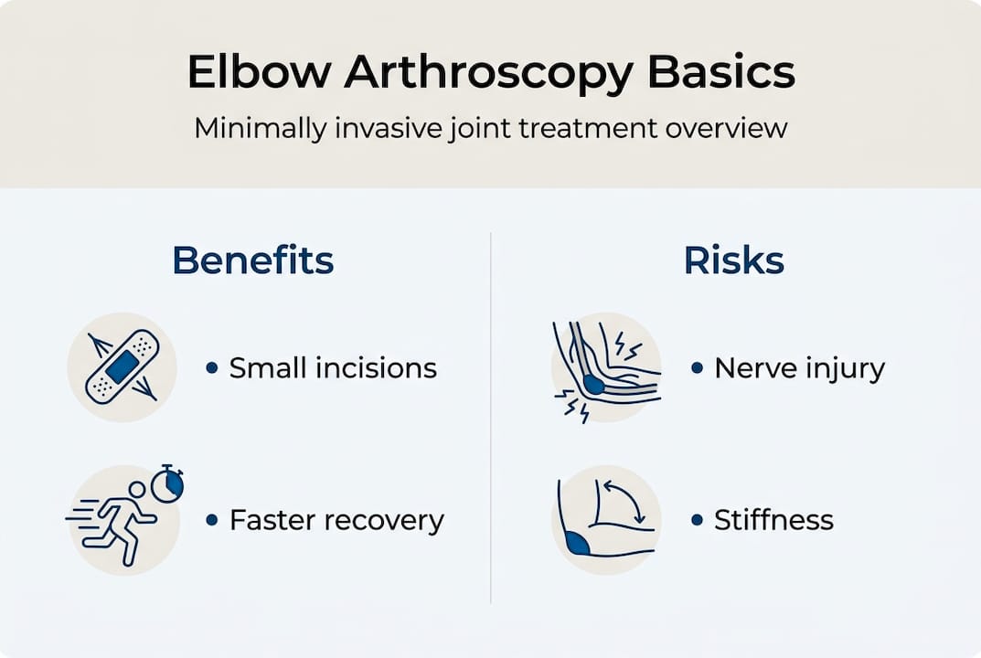
| Faster recovery | Compared with open surgery, elbow arthroscopy generally offers faster recovery and less postoperative pain. |
| Nerve safety emphasis | The elbow contains critical nerves near common portal sites, so choosing an experienced surgeon with extensive arthroscopic training reduces the risk of nerve injury. |
| Early rehabilitation | Starting physical therapy early after surgery improves range of motion and speeds overall recovery. |
What is elbow arthroscopy and how does it work?
Elbow arthroscopy is a minimally invasive surgical technique where an orthopedic surgeon inserts a thin, pencil-sized instrument called an arthroscope through small incisions to visualize, diagnose, and treat various elbow conditions. Unlike traditional open surgery requiring large incisions and extensive muscle dissection, arthroscopy treats conditions including osteoarthritis, rheumatoid arthritis, loose cartilage or bone fragments, elbow contractures limiting motion, osteochondral defects, and sports-related injuries with significantly less tissue trauma. The procedure typically takes place in an outpatient surgical center under general anesthesia or regional nerve blocks, allowing you to return home the same day.
The arthroscope contains a fiberoptic camera system that projects magnified images of your elbow joint onto a monitor, giving your surgeon a detailed view of internal structures. Through additional small portals, specialized miniaturized instruments like shavers, graspers, and radiofrequency probes are inserted to perform the necessary repairs or removals. The elbow surgeon can remove inflamed synovial tissue, extract loose bodies causing pain and catching, smooth damaged cartilage, release tight capsular tissue restricting motion, and repair ligaments or tendons depending on your specific condition.
The primary advantage of this approach lies in the incision size. Each portal measures approximately 1.5 centimeters compared to the 10 to 15 centimeter incisions required for open elbow surgery. Smaller incisions mean less pain, reduced scarring, lower infection risk, and faster healing times. You’ll experience less postoperative discomfort and typically require fewer pain medications than patients undergoing traditional open procedures.
Pro Tip: The elbow joint has limited space and critical nerves running close to common portal sites, so choosing a surgeon with extensive arthroscopic experience is essential for optimal safety and outcomes. Ask potential surgeons about their specific training in elbow arthroscopy and how many procedures they perform annually.
Common conditions treated with elbow arthroscopy:
- Degenerative or inflammatory arthritis causing pain and stiffness
- Loose bodies (bone or cartilage fragments) creating catching or locking sensations
- Post-traumatic contractures limiting elbow extension or flexion
- Osteochondritis dissecans and other cartilage injuries
- Tennis elbow or golfer’s elbow when conservative treatment fails
- Synovitis requiring tissue removal to reduce inflammation
Techniques and procedural considerations in elbow arthroscopy
Successful elbow arthroscopy requires meticulous attention to patient positioning, portal placement, and surgical technique due to the joint’s compact anatomy and proximity to neurovascular structures. Surgeons use three primary patient positions, each offering distinct advantages for visualization and access. The lateral decubitus position places you on your side with the affected arm supported, providing excellent access to posterior structures. The prone position has you lying face down with your arm hanging over a padded support, offering superior visualization of the posterior compartment and olecranon fossa. The supine position keeps you on your back with your arm across your chest or on an arm board, facilitating anterior and anterolateral access.
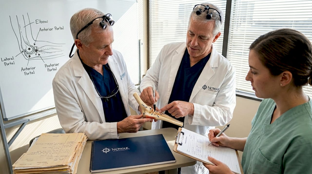
Portal placement represents the most critical safety consideration in elbow arthroscopy. Surgeons establish specific entry points to access different joint compartments while avoiding nerves and blood vessels. The anterolateral portal provides access to the radiocapitellar joint and anterior capsule. The mid-lateral portal offers excellent visualization of the central compartment. The posterolateral portal allows treatment of posterior loose bodies and synovitis. Each location carries specific nerve injury risks that experienced surgeons mitigate through precise anatomical knowledge and careful technique.
| Portal Location | Primary Access | Nerve at Risk | Best Positioning |
|---|---|---|---|
| Anterolateral | Radiocapitellar joint, anterior capsule | Radial nerve | Supine or lateral |
| Mid-lateral | Central compartment, capitellum | Posterior interosseous nerve | Any position |
| Posterolateral | Olecranon fossa, posterior compartment | Ulnar nerve | Prone or lateral |
| Anteromedial | Coronoid, medial compartment | Median nerve, brachial artery | Supine (use cautiously) |
Step-by-step approach to safe portal placement:
- Mark all bony landmarks and anticipated portal sites with the elbow flexed to 90 degrees before starting
- Distend the joint with sterile saline to push neurovascular structures away from the capsule
- Make skin incisions only, then use blunt dissection through subcutaneous tissue to the capsule
- Insert the arthroscope under direct visualization, never blindly advancing instruments
- Establish additional portals under arthroscopic guidance to ensure safe trajectories
- Continuously monitor instrument position relative to known neurovascular anatomy
Once portals are established, your surgeon performs the planned procedures. Joint distension with saline increases the working space and pushes nerves away from the surgical field. Debridement removes damaged cartilage and inflamed tissue using motorized shavers. Synovectomy excises thickened synovial lining contributing to pain and stiffness. Loose body removal extracts bone or cartilage fragments causing mechanical symptoms. Contracture release cuts tight capsular tissue to restore range of motion. The joint procedure complexity varies based on your specific pathology and treatment goals.
Pro Tip: The radial nerve crosses directly over the radiocapitellar joint just 3 millimeters from common portal sites. Surgeons who flex your elbow during portal creation and use proper distension techniques significantly reduce the already low risk of nerve injury.
Outcomes, risks, and recovery after elbow arthroscopy
Elbow arthroscopy delivers impressive results for appropriately selected patients. Studies show high success rates with Mayo Elbow Performance Scores ranging from 70 to 100 points, indicating good to excellent function. Approximately 77% of patients regain functional range of motion, with many achieving near-normal elbow extension and flexion. The typical procedure lasts one to two hours, and most patients go home the same day once anesthesia wears off and initial recovery monitoring is complete.
| Outcome Measure | Elbow Arthroscopy | Open Surgery |
|---|---|---|
| Average procedure time | 1-2 hours | 2-3 hours |
| Hospital stay | Same-day discharge | 1-2 days typical |
| Return to light activities | 2-4 weeks | 6-8 weeks |
| Full recovery timeline | 3-6 months | 6-12 months |
| Postoperative pain level | Mild to moderate | Moderate to severe |
| Complication rate | 5-11% | 8-15% |
While elbow arthroscopy is generally safe, complications can occur. Nerve injuries represent the most significant concern, particularly to the radial nerve which lies close to standard portal sites. Most nerve injuries are temporary neurapraxias that resolve within weeks to months, but permanent damage occasionally occurs. Infection rates remain low, typically under 1%, but any surgical procedure carries this risk. Postoperative stiffness can develop if you don’t begin physical therapy promptly or if excessive scar tissue forms. Persistent pain, though uncommon, affects some patients despite technically successful procedures.
“The radial nerve is at particular risk during elbow arthroscopy due to its superficial location near the radiocapitellar joint. Complication rates range from 5% to 11%, with nerve injury being the most concerning. Surgeon experience and meticulous technique are paramount to minimizing these risks.”
Common complications and warning signs:
- Nerve injury: numbness, tingling, weakness in hand or forearm (report immediately)
- Infection: increasing pain, redness, warmth, drainage, fever above 101°F
- Excessive stiffness: inability to improve range of motion despite therapy
- Persistent swelling: joint effusion lasting beyond 4-6 weeks
- Instrument breakage: rare but requires additional surgery if fragments remain
The good news is that arthroscopy provides outcomes comparable to open surgery for range of motion and functional improvement while offering faster rehabilitation and less postoperative pain. You’ll likely need fewer pain medications and return to daily activities weeks earlier than with traditional approaches. The cosmetic result is also superior, with small scars instead of long incisions.
Recovery follows a predictable timeline when you adhere to your rehabilitation program. The first week focuses on pain control, wound care, and gentle motion exercises. Weeks two through six emphasize progressive range of motion and light strengthening. Months two through four introduce functional activities and sport-specific training for athletes. Full recovery typically takes three to six months depending on the procedure complexity and your individual healing.
Early physical therapy plays a crucial role in optimizing outcomes. Starting gentle motion exercises within days of surgery prevents stiffness while promoting healing. Your therapist will guide you through progressive exercises designed to restore flexibility, rebuild strength, and retrain neuromuscular control. Compliance with your therapy program directly correlates with your final outcome, so prioritize these appointments even when progress feels slow.
Special cases and practical considerations for patients and athletes
Certain conditions require special consideration when evaluating elbow arthroscopy as a treatment option. Septic arthritis, or joint infection, traditionally required open surgical drainage, but arthroscopic treatment now offers a less invasive alternative for selected cases. Studies show good outcomes with arthroscopic irrigation and debridement, though complication rates run slightly higher than for routine procedures. Your surgeon must weigh the benefits of smaller incisions against the need for thorough infection clearance when infection is present.
Post-traumatic elbow stiffness presents another challenging scenario. After fractures or dislocations, scar tissue and bone spurs can severely limit motion. Some surgeons prefer open capsular release and heterotopic ossification removal for maximum visualization and bone work. Others advocate for arthroscopic approaches to minimize additional trauma. The debate continues, with outcomes depending heavily on surgeon experience and the specific pathology present. If you’re dealing with post-traumatic stiffness, discuss both options thoroughly with your orthopedic specialist.
Preoperative imaging is essential for surgical planning and diagnosis. MRI scans reveal soft tissue pathology including ligament tears, cartilage damage, and synovitis that X-rays miss. Standard X-rays show bone spurs, loose bodies, and arthritis. Your surgeon reviews these images to map portal placement, anticipate anatomical variations, and plan the specific procedures needed to address your condition. Never skip recommended imaging studies, as they directly impact surgical safety and effectiveness.
Athletes face unique considerations when contemplating elbow arthroscopy. Overhead throwing athletes, tennis players, and gymnasts place enormous stress on elbow structures and demand complete restoration of function. Your surgeon should use sport-specific outcome measures and understand the biomechanical demands of your activity. Return to sport protocols must be individualized, with throwing athletes often requiring four to six months before resuming competitive pitching even after successful surgery.
Key rehabilitation strategies for optimal recovery:
- Begin gentle passive range of motion exercises within 48-72 hours post-surgery
- Progress to active-assisted motion by week two, avoiding forceful stretching
- Introduce light resistance training at weeks four to six once motion improves
- Incorporate proprioceptive and neuromuscular training by month two
- Return to sport-specific activities only after achieving full motion and 85% strength
- Maintain long-term flexibility and strengthening programs to prevent recurrence
Pro Tip: If you’re an athlete or highly active adult, seek surgeons who specialize in sports injuries and regularly treat patients with similar demands. Ask about their return-to-sport rates and timelines for your specific activity. Starting early, supervised rehabilitation with a therapist experienced in athletic recovery dramatically improves your chances of returning to pre-injury performance levels.
The success of elbow arthroscopy in complex cases like osteoarthritis management depends on realistic expectations. While the procedure effectively removes loose bodies, smooths rough cartilage, and releases contractures, it cannot reverse advanced joint degeneration. You may experience significant symptom relief and improved function, but the underlying arthritis will progress over time. Discuss long-term prognosis honestly with your surgeon to ensure arthroscopy aligns with your goals.
Explore expert orthopedic care at Nowak Orthopedic Associates
If you’re struggling with elbow pain or have been told you need surgery, exploring minimally invasive options with an experienced specialist can make all the difference in your recovery. Nowak Orthopedic Associates offers comprehensive evaluation and treatment for elbow injuries and conditions, combining advanced arthroscopic techniques with a multidisciplinary approach to healing. Dr. Nowak’s expertise in sports injury treatment ensures athletes and active adults receive personalized care designed to restore function and get you back to your activities safely. Beyond surgery, the practice provides cutting-edge orthobiologic injections including PRP therapy to enhance healing and potentially avoid surgical intervention. Discover the full range of orthopedic services available and schedule a consultation to discuss your specific condition and treatment options.
FAQ
How painful is elbow arthroscopy?
Elbow arthroscopy uses small incisions and either regional nerve blocks or general anesthesia, which means you feel no pain during the actual procedure. Postoperative discomfort is typically less than open surgery because there’s minimal muscle and tissue disruption. Most patients describe the pain as moderate and manageable with prescribed medications for the first few days, then quickly subsiding to mild soreness. Ice, elevation, and following your surgeon’s pain management protocol help control discomfort during the initial recovery period.
What is the typical recovery time after elbow arthroscopy?
Most patients go home the same day as surgery once they’ve recovered from anesthesia and demonstrated adequate pain control. You’ll notice gradual improvement over the following weeks, with significant gains in the first month as swelling decreases and motion returns. Full recovery typically takes three to six months depending on the complexity of your procedure and how diligently you follow your rehabilitation program. Early physical therapy starting within the first week is essential for restoring range of motion efficiently and preventing stiffness that could prolong recovery.
Are there risks unique to elbow arthroscopy compared to open surgery?
Nerve injury risk is slightly higher with arthroscopy due to the tight joint space and proximity of the radial, ulnar, and median nerves to standard portal sites. The radial nerve is particularly vulnerable as it crosses just millimeters from common entry points. However, surgeons who use proper distension techniques, careful portal placement, and meticulous surgical methods keep this risk very low, typically under 5%. Infection and stiffness risks exist with both approaches at similar rates, generally remaining below 2% for infection and 5-10% for significant postoperative stiffness.
Who is an ideal candidate for elbow arthroscopy?
Athletes and active adults experiencing elbow pain from arthritis, loose bodies, contractures, or sports injuries often benefit most from arthroscopic treatment. You’re a good candidate if conservative treatments like physical therapy, medications, and injections haven’t provided adequate relief and imaging studies confirm pathology amenable to arthroscopic repair. Your surgeon will evaluate you with a thorough physical examination, review X-rays and MRI scans, and discuss your activity goals before recommending arthroscopy. Patients with realistic expectations, good overall health, and commitment to postoperative rehabilitation achieve the best outcomes.