

TL;DR:
- Elbow instability involves abnormal, painful joint movement caused by ligament or structural damage.
- It manifests in various types like posterolateral rotatory, valgus, and chronic instability, often linked to trauma or overuse.
- Accurate diagnosis requires dynamic testing and expert evaluation for effective nonoperative or surgical treatment.
Many people spend months managing what they believe is a strained elbow, only to discover the real problem is something more complex. Elbow instability, which is excessive painful joint mobility often caused by trauma, overuse, or connective tissue disease, is frequently overlooked in early assessments. This article explains what elbow instability actually is, the forms it takes, why it happens, how specialists diagnose it, and what treatment options can genuinely restore your quality of life.
Table of Contents
- What is elbow instability?
- Types and patterns of elbow instability
- Mechanics and risk factors: Why instability happens
- Diagnosis and evaluation pathways
- Treatment strategies: Nonoperative and surgical options
- A fresh perspective: What most people (and even some providers) miss about elbow instability
- Next steps: Get expert orthopedic help for elbow instability
- Frequently asked questions
Key Takeaways
| Point | Details |
|---|---|
| Elbow instability is distinct | Instability involves painful and abnormal joint movement, not just general soreness. |
| Multiple patterns exist | Elbow instability has different causes and patterns, with PLRI the most common. |
| Diagnosis requires expertise | Accurate evaluation uses advanced tests and imaging for reliable diagnosis. |
| Treatment is effective | Both bracing and surgical repair offer high rates of return to normal activity and pain relief. |
What is elbow instability?
Elbow instability is not simply pain in the joint. It refers specifically to abnormal movement within the elbow that the surrounding structures can no longer control. According to orthopedic specialists, elbow instability involves excessive and usually painful mobility in the elbow joint, most often resulting from trauma, overuse, or connective tissue disease. That distinction matters because it changes everything about how the condition must be evaluated and treated.
Your elbow is a hinge joint stabilized by a network of ligaments, tendons, and bony anatomy. When any part of that network is compromised, the joint can shift in ways it shouldn’t. That abnormal motion creates pain, of course, but it also leads to functional limitations that affect daily tasks like lifting, pushing, or throwing.
Common causes of elbow instability include:
- Acute trauma: A fall on an outstretched hand, a direct blow, or a sports collision can partially or fully dislocate the elbow and tear stabilizing ligaments.
- Overuse injury: Repetitive stress from throwing sports, racket sports, or manual labor gradually weakens ligament integrity over time.
- Prior elbow surgery or fracture: Structural changes after a previous injury can compromise the mechanics of the joint.
- Connective tissue disorders: Conditions that affect collagen or ligament quality can predispose patients to instability even without major trauma.
Elbow instability is different from arthritis or tendinitis. It’s a structural problem with the joint’s ability to stay properly aligned during movement.
Consulting with experienced elbow orthopedic surgeons is critical because the symptoms can mimic other elbow conditions, and general practitioners may not order the specialized testing needed to identify instability. Patients often notice their elbow feels like it wants to give out, clicks unexpectedly, or simply doesn’t feel right even after initial treatment for a supposed strain. If that sounds familiar, exploring elbow pain relief options with a specialist is an important step forward.
Types and patterns of elbow instability
With the basics understood, it’s important to see the many forms elbow instability can take, because the type directly influences how it is diagnosed and managed.
Elbow instability can be classified by timing, the joint involved, the degree and direction of displacement, and associated fractures. The most common types include posterolateral rotatory instability (PLRI), valgus instability, varus instability, posteromedial rotatory instability, and anterior instability (which is rare).
Here is a brief comparison of the main types:
| Type | Key feature | Most common in |
|---|---|---|
| PLRI | Abnormal rotatory subluxation | Trauma, post-surgical patients |
| Valgus instability | Medial ligament failure | Overhead throwers, pitchers |
| Varus instability | Lateral ligament failure | Contact sport athletes |
| Posteromedial rotatory | Coronoid stress fractures | Middle-aged adults |
| Anterior (rare) | Anterior displacement | High-energy injury |
Instability also follows a general timeline that helps orthopedic specialists categorize severity:
- Acute instability: Occurs immediately after injury, usually accompanied by swelling, sharp pain, and obvious dysfunction.
- Subacute instability: Develops in the weeks following an injury that was partially treated but not fully resolved.
- Chronic instability: Persists for months or years, often becoming the patient’s new normal because early diagnosis was missed.
- Recurrent instability: Episodes of giving way or subluxation (partial dislocation) that return repeatedly after apparent recovery.
Understanding the pattern of instability is also useful when thinking about related joint procedure examples and the surgical or nonsurgical interventions that may be appropriate.
Pro Tip: Many cases of chronic instability are missed on routine X-rays taken at rest. Dynamic stress testing, where the joint is loaded under movement, is often essential to capture instability that static images simply cannot show.
Mechanics and risk factors: Why instability happens
Understanding the patterns is useful, but let’s look at what’s happening inside the joint and who is most at risk.
The elbow relies on several ligament complexes for stability. The lateral ulnar collateral ligament (LUCL) on the outer side controls rotatory motion. The medial ulnar collateral ligament (MUCL) on the inner side resists the valgus force (outward bending) that occurs during overhead throwing. When either structure is injured or becomes chronically stretched, the joint loses its ability to stay centered during movement.

PLRI specifically involves abnormal rotatory subluxation caused by LUCL injury, while the terrible triad injury refers to a dislocation combined with radial head fracture and coronoid fracture resulting from a specific high-energy force pattern. The terrible triad is one of the most surgically complex elbow injuries because it compromises both ligamentous and bony stability simultaneously.
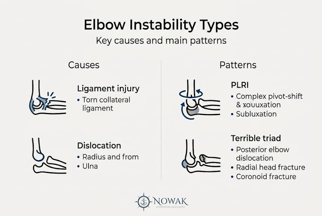
Risk factors vary depending on the type of instability:
| Risk factor | Associated instability type |
|---|---|
| Overhead throwing sports | Valgus (MUCL injury) |
| Falls on outstretched arm | PLRI or acute dislocation |
| Prior elbow surgery/fracture | Post-surgical instability |
| Contact sports | Varus, acute trauma |
| Hypermobility syndromes | Multiple types, often bilateral |
Athletes who throw, including baseball pitchers, quarterbacks, javelin throwers, and tennis players, place extraordinary valgus stress on the medial elbow with every repetition. Over a career, this cumulative load can cause the MUCL to progressively loosen, leading to valgus instability that severely limits performance before it causes obvious structural failure.
Patients who have previously dislocated their elbow and returned to activity without proper ligament healing are especially vulnerable to developing recurrent or chronic instability. The athlete injury workflow used by orthopedic specialists in sports medicine addresses this risk with targeted return-to-play protocols.
Diagnosis and evaluation pathways
Recognizing symptoms is only the start. How do orthopedic experts make the diagnosis definitive?
The diagnostic process for elbow instability follows a careful, stepwise approach. Diagnosis involves history, physical examination using specific provocation tests, and imaging studies including X-ray, MRI, CT, and sometimes fluoroscopy (real-time moving X-ray).
Here is a typical evaluation sequence:
- Detailed symptom history: When did symptoms start, what makes them worse, and has there been prior trauma or surgery?
- Physical examination: Specialized tests like the lateral pivot shift test, the valgus stress test, and the posterolateral drawer test place controlled load on the elbow to reproduce instability.
- Standard X-rays: Useful for identifying fractures or significant malalignment, but often appear normal in isolated ligament instability.
- MRI: The preferred imaging tool for assessing ligament integrity, soft tissue swelling, and associated cartilage damage.
- CT scan: Particularly valuable when bony involvement or fracture pattern needs precise evaluation before surgery.
- Dynamic fluoroscopy or stress imaging: Captures instability during movement when static images are inconclusive.
Seeing a dedicated elbow arthroscopy guide can also help patients understand how minimally invasive diagnostic arthroscopy is sometimes used to directly visualize the joint when imaging remains unclear.
Pro Tip: If your elbow persistently locks, snaps, or gives out after a known injury, even if X-rays came back normal, request an MRI or a referral to an orthopedic specialist for advanced evaluation. Normal X-rays do not rule out instability.
Treatment strategies: Nonoperative and surgical options
A clear diagnosis leads directly to creating a personalized treatment plan, and the good news is that most patients have meaningful options regardless of severity.
For acute or straightforward cases, nonoperative management is typically the starting point. This involves reduction (realigning the joint if dislocated), early controlled motion to prevent stiffness, and protective bracing that allows healing while limiting harmful movement. Physical therapy focuses on strengthening the muscles around the elbow to compensate for ligament weakness during the recovery phase.
Surgical intervention becomes necessary when instability is severe, recurrent, or fails to respond to conservative care. The primary surgical option for PLRI is repair or reconstruction of the LUCL, restoring the joint’s rotational stability. For athletes with valgus instability, MUCL reconstruction (commonly known as Tommy John surgery) is highly effective.
Research shows that LUCL reconstruction produces good to excellent outcomes in 85 to 90% of patients. That is a strong success rate that gives patients real reason for confidence when surgery is recommended.
Key recovery milestones for most patients include:
- Weeks 1 to 3: Pain and swelling management, gentle range-of-motion exercises.
- Weeks 4 to 8: Progressive strengthening and functional movement training.
- Months 3 to 4: Return to light sport-specific activity for most non-throwing athletes.
- Months 9 to 12: Full return to overhead throwing or contact sports after ligament reconstruction.
For a full breakdown of available procedures, the elbow surgery options available at our practice cover everything from conservative arthroscopic repair to complex ligament reconstruction.
A fresh perspective: What most people (and even some providers) miss about elbow instability
Beyond protocols and imaging checklists, clinical experience reveals a pattern that many patients and providers miss: PLRI is frequently underdiagnosed because standard static X-rays appear completely normal, and the subtle giving-way sensation patients describe is too often attributed to vague “pain” or prior injury.
The uncomfortable reality is that patients with chronic elbow instability sometimes see multiple providers over months or even years before receiving the correct diagnosis. The elbow is simply not evaluated with the same instinctive suspicion that, say, the knee or shoulder receives after trauma.
Our strong recommendation: if your elbow symptoms have persisted beyond six to eight weeks despite treatment, seek an orthopedic specialist who routinely evaluates instability with dynamic testing, not just routine imaging. Timely diagnosis is the single factor most strongly associated with better outcomes.
Next steps: Get expert orthopedic help for elbow instability
If you suspect instability, connecting with experts is your best next step. At Nowak Orthopedic Associates, we provide orthopedic joint care that goes beyond routine evaluations. Dr. Nowak applies advanced diagnostic tools and personalized treatment planning to identify and address the true source of your elbow problems. Whether your path forward involves bracing, therapy, regenerative treatments, or minimally invasive elbow treatment, we are ready to guide you. Schedule your first visit and take a confident step toward lasting relief.
Frequently asked questions
What are the warning signs of elbow instability?
Common signs include pain with movement, a locking or catching sensation, the feeling that your elbow is giving out, and difficulty with activities like gripping or lifting. These symptoms, including snapping and a sense of giving way, should prompt evaluation by an orthopedic specialist.
Is surgery always needed for elbow instability?
No. Nonoperative management is preferred for most acute or simple cases, with surgical intervention reserved for complex, severe, or recurrent instability that does not respond to bracing and therapy.
How successful is treatment for elbow instability?
Both nonoperative and surgical treatments carry high success rates. PLRI reconstruction achieves good or excellent outcomes in 85 to 90% of patients, giving most people strong confidence in pursuing appropriate care.
Can athletes return to their sport after elbow instability treatment?
Yes. Most athletes achieve high return-to-activity rates after completing rehabilitation and, when needed, undergoing ligament reconstruction surgery, with many returning to full competitive participation.