

TL;DR:
- Cartilage cannot repair itself naturally because it lacks a blood supply, requiring surgical intervention. OATS uses your own healthy cartilage and bone from non-weight-bearing areas to restore small to medium-sized defects with hyaline cartilage for durable joint repair. Proper patient selection, age, defect size, and commitment to recovery are essential for successful outcomes with OATS surgery.
Cartilage damage is one of the most frustrating orthopedic problems you can face. Unlike bone, cartilage has almost no blood supply, which means it cannot repair itself after injury. If you have been told you have a cartilage defect in your knee, you have probably heard that your options are limited. That is where understanding what is OATS cartilage restoration becomes genuinely useful. OATS, short for osteochondral autograft transfer system, is a surgical technique that moves healthy cartilage from one part of your joint to the damaged area. This guide covers how the procedure works, who qualifies, what recovery looks like, and how it compares to other cartilage repair options.
Table of Contents
- Key takeaways
- What is OATS cartilage restoration?
- Who is a good candidate for OATS?
- What recovery looks like after OATS surgery
- Comparing OATS to other cartilage repair options
- My perspective on OATS and patient expectations
- How Nowakorthopedics can help you move forward
- FAQ
Key takeaways
| Point | Details |
|---|---|
| Cartilage cannot self-repair | Damaged joint cartilage requires surgical intervention because it lacks a blood supply to heal naturally. |
| OATS uses your own tissue | Healthy cartilage is harvested from a non-weight-bearing area of your own joint and transplanted to the defect. |
| Best for focal, smaller defects | OATS works best for small to medium-sized cartilage lesions in weight-bearing areas of the knee. |
| Recovery takes several months | Full return to activity typically takes six months or more, with structured physical therapy playing a major role. |
| Multiple repair options exist | OATS is one of several cartilage restoration techniques, and the right choice depends on defect size, age, and activity level. |
What is OATS cartilage restoration?
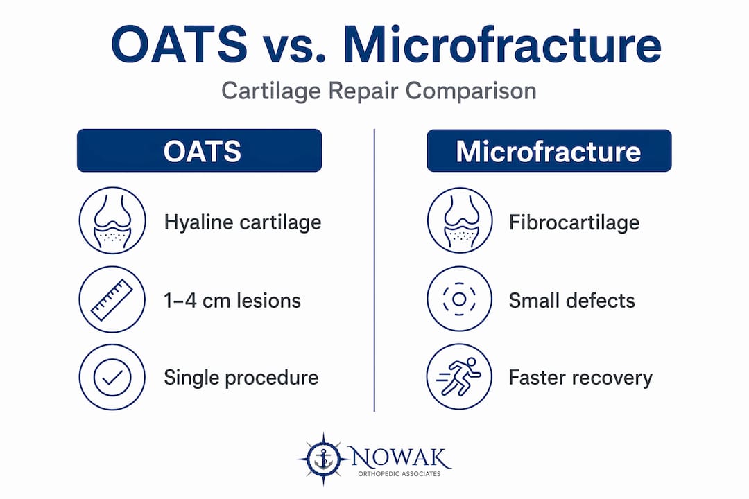
OATS stands for osteochondral autograft transfer system. The procedure involves harvesting healthy cartilage and underlying bone from a non-weight-bearing region of your joint, then transplanting that plug into the damaged cartilage site. The graft contains what is called hyaline cartilage, which is the same high-quality cartilage that lines your joint surfaces. This is a meaningful distinction because other repair methods often produce fibrocartilage, a scar-like tissue that is structurally inferior and tends to break down faster over time.
The surgeon typically performs OATS either arthroscopically or through a small open incision, depending on the size and location of the defect. A cylindrical plug of bone and cartilage is removed using a specialized coring tool, and a matching plug is then press-fit into the prepared defect site. The bone portion integrates with the surrounding bone over time, anchoring the cartilage graft securely. Bone and cartilage growth into the damaged area leads to restored joint function and sustained pain relief.
When the damaged area is larger, a surgeon may use multiple plugs placed in a mosaic pattern. This variation is known as mosaicplasty. Both OATS and mosaicplasty use osteochondral cores from non-weight-bearing areas, but mosaicplasty uses several smaller grafts rather than one or two larger ones. The choice between them depends on defect geometry and surgeon preference.
Pro Tip: Ask your surgeon specifically whether your procedure will be performed arthroscopically or open, and what size graft is planned. These details directly affect your recovery timeline and weight-bearing restrictions.
Here is a quick comparison of how OATS relates to similar cartilage restoration techniques:
| Feature | OATS | Mosaicplasty |
|---|---|---|
| Graft type | Autograft (own tissue) | Autograft (own tissue) |
| Number of plugs | Usually 1 to 2 | Multiple small plugs |
| Best defect size | Small to medium | Small to medium |
| Cartilage quality | Hyaline | Hyaline |
| Coverage consistency | High for single lesion | Variable fill between plugs |
Who is a good candidate for OATS?
OATS is mainly performed for focal osteochondral defects of the knee joint, particularly in weight-bearing regions like the femoral condyle. Patients who benefit most tend to share a specific set of characteristics.
Good candidates typically include:
- Younger to middle-aged patients (generally under 50) with otherwise healthy joint cartilage
- Individuals with isolated, focal cartilage defects rather than widespread joint degeneration
- Active patients who want to return to sports or physically demanding activities
- Those diagnosed with osteochondritis dissecans, a condition where a fragment of bone and cartilage separates from the joint surface
The size of the defect matters considerably. OATS performs best for lesions in the range of one to four square centimeters. Larger defects may require alternative techniques such as autologous chondrocyte implantation (ACI) or osteochondral allograft transplantation.
There are also important limitations to recognize. Patients with widespread osteoarthritis of the knee are generally not candidates because the surrounding cartilage is already compromised. OATS is also considered investigational for certain locations, such as the patellar surface or distal femoral articular regions outside of specific indications. Your surgeon will use imaging, including MRI, to assess defect size, depth, and surrounding tissue health before recommending the procedure.
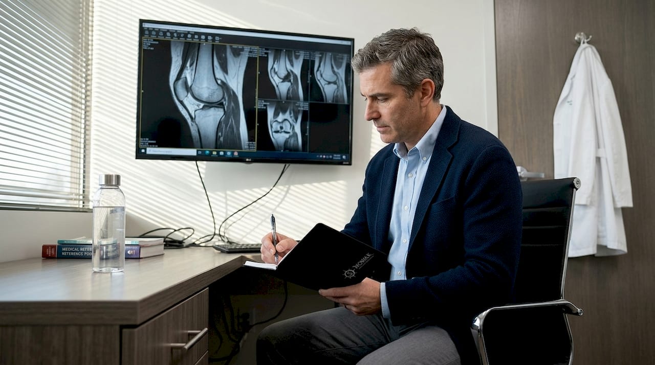
Pro Tip: If you are unsure whether your cartilage damage qualifies for OATS, ask for an MRI with cartilage-sensitive sequencing. A standard X-ray will not show cartilage defects clearly.
Evidence supports strong outcomes for appropriate candidates. Among adolescents treated with OATS for osteochondritis dissecans, the return to sport rate reached 95%, with an average return time of around six months.
What recovery looks like after OATS surgery
Recovery from OATS surgery is structured and gradual. Rushing it is the most common mistake patients make, and it directly affects long-term outcomes. Here is what a typical recovery progression looks like:
- Weeks 1 to 6: Protected weight bearing with crutches. The focus is on controlling swelling, managing pain, and maintaining range of motion through gentle exercises.
- Weeks 6 to 12: Progressive weight bearing as tolerated. Physical therapy intensifies with strengthening exercises targeting the quadriceps and surrounding muscles.
- Months 3 to 5: Functional training begins. Low-impact activities like cycling and swimming are usually introduced during this phase.
- Months 5 to 6 and beyond: Sport-specific training and gradual return to full activity, contingent on achieving strength and stability benchmarks.
Postoperative rehabilitation after OATS focuses on restoring motion, building strength, and returning to activity over several months. Skipping or reducing physical therapy during this period significantly reduces the chance of a full recovery.
Pain management early on typically involves ice, elevation, and prescribed medications. Many patients are surprised by how manageable discomfort is after the first week, but swelling can persist for several months as the graft integrates.
Pro Tip: Stick to your physical therapy schedule even when you feel good. Cartilage graft integration continues for months after surgery, and premature stress on the joint can compromise the repair.
Comparing OATS to other cartilage repair options
Understanding OATS surgery means understanding how it fits within the broader menu of cartilage repair options. Each technique has specific indications, and no single method works for everyone.
| Procedure | Graft Source | Best Defect Size | Recovery Time | Relative Cost |
|---|---|---|---|---|
| OATS | Patient’s own tissue | Small to medium | 4 to 6 months | Moderate (~$6,110) |
| Microfracture | None (bone marrow stimulation) | Small | 3 to 4 months | Lower |
| ACI / MACI | Patient’s own cells (lab grown) | Medium to large | 9 to 18 months | Higher |
| Allograft | Donor tissue | Large | 6 to 12 months | Moderate to high |
Microfracture is the simplest option and works by drilling tiny holes into bone beneath the defect to stimulate marrow cells. The tissue it produces is fibrocartilage rather than hyaline cartilage, which tends to degrade faster. It works reasonably well for small defects in younger patients, but outcomes decline for lesions over two square centimeters.
ACI and its matrix-assisted variant (MACI) involve harvesting your own cartilage cells, expanding them in a lab, and reimplanting them. Clinical scores with MACI remained improved over 20 years, making it one of the most durable options for larger defects. However, it requires two surgeries, a longer recovery, and significantly higher cost.
OATS stands out because it delivers hyaline cartilage in a single procedure using the patient’s own tissue. Perioperative costs average around $6,110, placing it between the lower cost of microfracture and the higher cost of ACI. For the right patient with a focal, medium-sized defect, it offers a strong balance of durability, recovery time, and cost.
My perspective on OATS and patient expectations
I have seen OATS produce genuinely life-changing results for the right patients. A young athlete with an isolated femoral condyle defect who commits fully to rehab can return to sport at a level many never expected possible.
That said, what I find patients underestimate most is how much the recovery phase determines the final outcome. The surgery itself is only half the equation. Patients who engage seriously with physical therapy, respect weight-bearing restrictions, and show up consistently to follow-up appointments do better. Those who push too fast almost always pay for it.
There is also a candidacy issue that does not get enough attention. Some patients come in hoping OATS will solve what is actually early-stage osteoarthritis. It will not. Offering OATS to a patient with diffuse joint degeneration does them no favors, and part of our job at Nowakorthopedics is making sure candidates are selected carefully using imaging and a thorough clinical assessment.
If you are considering OATS, be honest with your surgeon about your activity goals and your ability to commit to recovery. The procedure rewards motivated patients and penalizes those who cut corners.
— Rich
How Nowakorthopedics can help you move forward
If you are dealing with knee cartilage damage and want to know whether OATS is right for you, Nowakorthopedics is here to help. Dr. Roger B. Nowak performs OATS cartilage restoration as part of a full range of knee joint preservation procedures designed to restore function and reduce pain. Every patient receives a personalized evaluation that considers defect size, activity level, and long-term goals before any surgical recommendation is made. Whether you need surgical repair or want to explore non-surgical alternatives first, the team at Nowakorthopedics will build a plan around your specific situation. You can learn more about what to expect by reviewing our guide to advanced joint care or schedule your first visit to get started today.
FAQ
What is OATS cartilage restoration in simple terms?
OATS is a surgical procedure that moves a plug of healthy cartilage and bone from a non-weight-bearing area of your joint to a damaged area, restoring a smooth, functional joint surface using your own tissue.
How long does recovery from OATS surgery take?
Most patients return to full activity within five to six months, though complete graft integration can continue for up to a year. Physical therapy throughout the recovery period is critical to achieving full function.
Is OATS better than microfracture?
For small to medium focal defects, OATS generally produces more durable results because it transplants hyaline cartilage rather than stimulating lower-quality fibrocartilage growth, as microfracture does.
How much does OATS surgery cost?
OATS surgery costs around $6,110 on average, making it more expensive than microfracture but less costly than autologous chondrocyte implantation. Costs vary based on facility, location, and insurance coverage.
Who is not a good candidate for OATS?
Patients with widespread osteoarthritis, large cartilage defects covering several joint surfaces, or significant bone loss may not be suitable for OATS. Certain locations on the knee, such as the patellar surface, may also fall outside standard OATS indications.