

TL;DR:
- Proper diagnosis of shoulder injuries relies on detailed history, physical examination, and selective imaging to avoid unnecessary treatments. Recognizing signs like weakness, deformity, and persistent pain guides timely intervention, while understanding the limitations of imaging ensures accurate clinical correlation. A thorough evaluation by a qualified specialist helps determine the appropriate treatment plan for optimal recovery.
Shoulder pain has a way of making everyday life unexpectedly difficult. Reaching for a coffee mug, sleeping on your side, or lifting a bag onto a shelf can all become uncomfortable reminders that something is wrong. Knowing how to diagnose shoulder injuries accurately matters because the wrong assumption leads to the wrong treatment. Whether you recently hurt yourself or have been dealing with a nagging ache for months, understanding the diagnostic process puts you in a much stronger position to get the right care and recover fully.
Table of Contents
- Key Takeaways
- How to diagnose shoulder injuries: start with preparation
- Physical examination and self-assessment tests
- Diagnostic imaging and when it is necessary
- Common mistakes in diagnosing shoulder injuries
- What to do after a shoulder injury diagnosis
- My perspective on clinical evaluation
- Get a thorough shoulder evaluation at Nowak Orthopedic Associates
- FAQ
Key Takeaways
| Point | Details |
|---|---|
| Start with patient history | Tracking when pain started and what makes it worse helps clinicians identify injury type faster. |
| Red flags need urgent care | Visible deformity, severe weakness, or numbness warrant immediate medical evaluation. |
| Physical exam comes first | Clinical tests for rotator cuff and impingement often guide diagnosis before any imaging is ordered. |
| Imaging has limits | X-rays show bone, not soft tissue. MRI or ultrasound is needed to confirm tears and tendon damage. |
| Early diagnosis prevents chronic damage | Ignoring weakness or clicking sounds delays treatment and raises the risk of long-term degeneration. |
How to diagnose shoulder injuries: start with preparation
Before any physical test or scan, the most useful diagnostic tool is your own account of what happened. Tracking mechanism of injury gives clinicians critical context. A sudden fall or collision raises suspicion for fractures and acute tears. Gradual onset over weeks or months points more toward chronic conditions like tendinopathy or arthritis.
When you speak with a healthcare provider, be prepared to describe the following:
- When the pain started and whether it came on suddenly or gradually
- What activities or positions make it worse or better
- Whether the pain is sharp and stabbing, a dull ache, or burning
- Whether you feel numbness or tingling down your arm
- What treatments you have already tried, including rest, ice, or over-the-counter medication
Certain symptoms require more than a scheduled appointment. Seek immediate attention when you notice visible deformity, severe and sudden weakness, chest pressure accompanied by shoulder pain, or numbness that does not go away. These can signal fractures, full tendon ruptures, or even cardiac events referred to the shoulder.
Pro Tip: Keep a short daily log of your shoulder symptoms for one week before your appointment. Note pain level, time of day, and what triggered it. This gives your provider a clearer picture than memory alone.
Physical examination and self-assessment tests
A structured shoulder injury assessment follows a logical sequence: look, move, test. Each step reveals different information.
Visual inspection comes first. You or a clinician looks for asymmetry between shoulders, visible swelling, muscle wasting (atrophy), or any obvious deformity. Noticeable muscle loss around the back of the shoulder often suggests a long-standing rotator cuff problem.
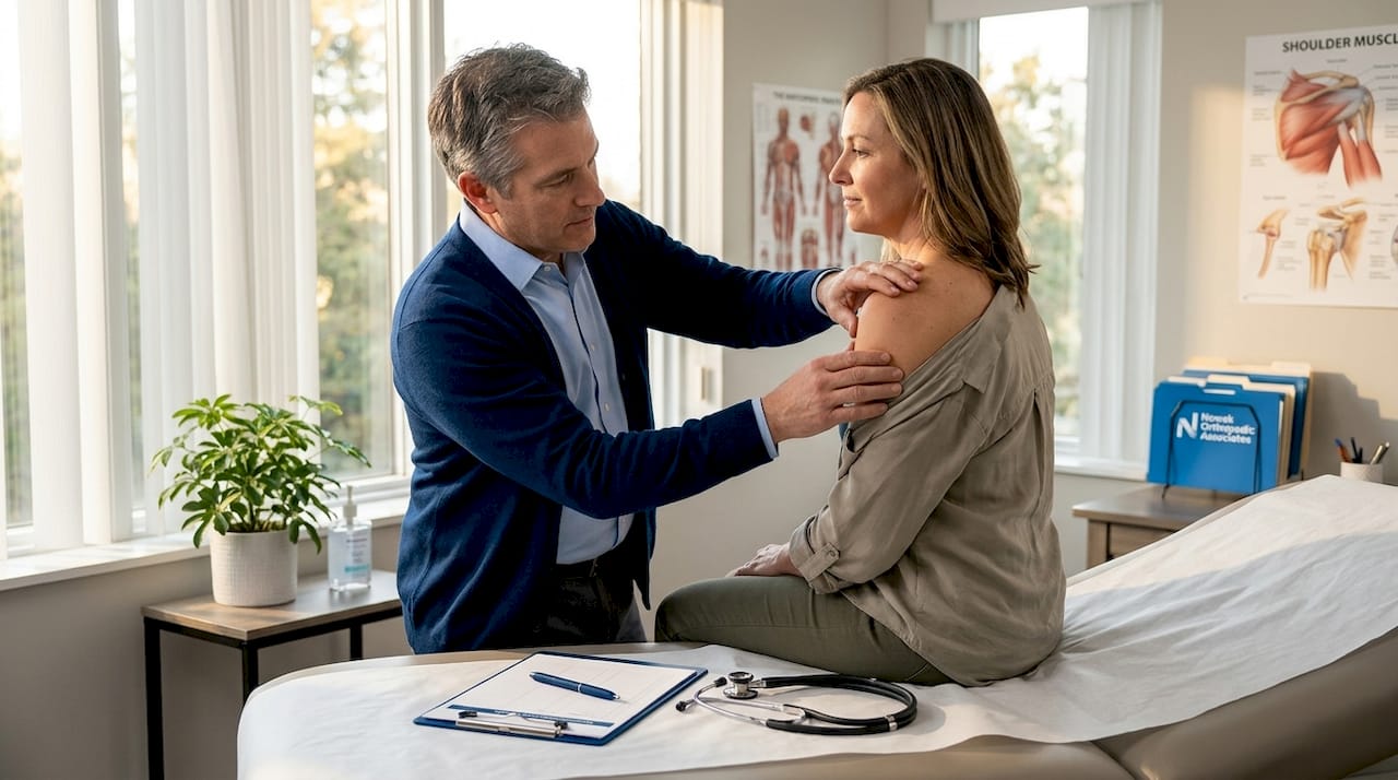
Range of motion testing comes next, and the distinction between active and passive motion matters. Active range of motion is what you can move on your own. Passive range of motion is how far someone else can move your arm while you relax. If passive motion is significantly better than active motion, that points to a muscle or tendon problem rather than a stiff joint.
Special orthopedic tests are maneuvers designed to stress specific structures. The Neer, Hawkins-Kennedy, and drop-arm tests are among the most commonly used in clinical settings. Here is a quick reference:
| Test | What it detects | How to interpret it |
|---|---|---|
| Neer test | Subacromial impingement | Pain on forward arm raise suggests impingement |
| Hawkins-Kennedy test | Rotator cuff impingement | Pain on internal rotation at 90° is a positive sign |
| Drop arm test | Full-thickness rotator cuff tear | Inability to hold arm at 90° suggests significant tear |
| Empty can test | Supraspinatus tendon integrity | Weakness or pain with resisted abduction is positive |
Research shows that inability to abduct above 90° has a diagnostic odds ratio of 12.9 for full-thickness rotator cuff tears, making it one of the most telling signs in a physical exam. Pain on resisted abduction carries 91% sensitivity for the same condition.
It is worth noting that many special tests have uncertain standalone accuracy and must be interpreted alongside the full clinical picture. No single maneuver confirms a diagnosis on its own.
Pro Tip: Do not push through significant pain during self-assessment. These tests are meant to detect symptoms, not force them. If a movement causes sharp pain, stop and note it rather than repeating it.
Diagnostic imaging and when it is necessary
Imaging is a powerful tool, but it is not always the first or most important step. Orthopedic specialists prioritize a quality physical exam precisely because imaging, especially MRI, frequently reveals incidental findings that have nothing to do with the patient’s actual pain.
Here is how the main imaging types are used:
| Imaging type | Best for | Limitations |
|---|---|---|
| X-ray | Fractures, arthritis, bone spurs | Cannot visualize tendons or soft tissue |
| MRI | Rotator cuff tears, labral injuries, bursitis | Expensive; incidental findings are common |
| Ultrasound | Tendon tears, dynamic tendon assessment | Operator-dependent; limited for deep structures |
X-rays assess bony anatomy and are usually the first imaging ordered. MRI and ultrasound are reserved for cases where soft tissue injury is suspected and the clinical picture is unclear. Ultrasound has the advantage of showing tendons in motion, which can be useful for evaluating certain conditions dynamically.
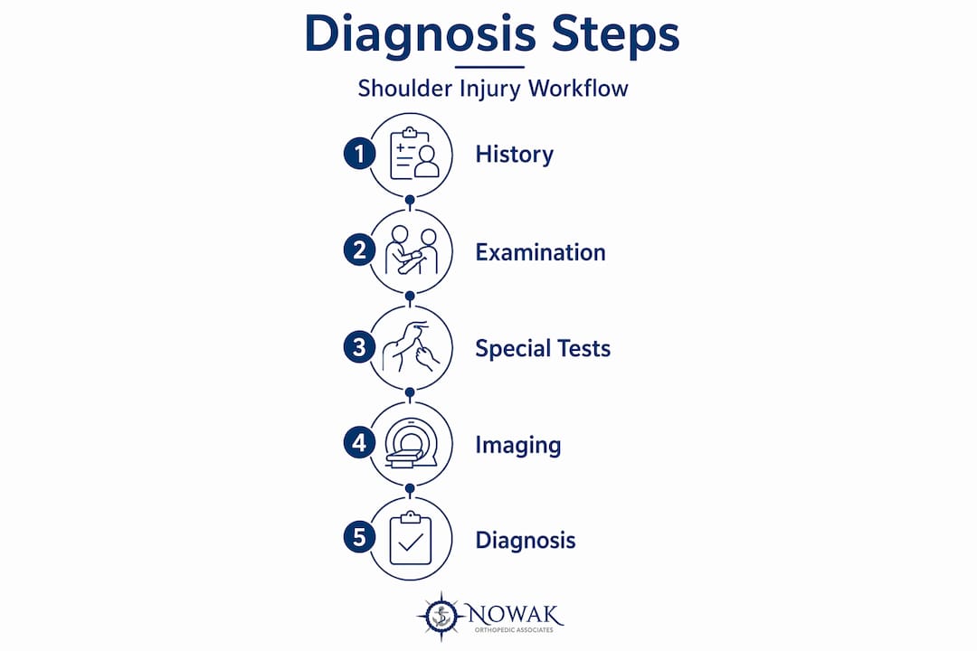
One of the most common pitfalls in shoulder pain diagnosis is over-relying on imaging results without connecting them to symptoms. A partial rotator cuff tear found on MRI in a patient with minimal pain may not be the source of their problem. Imaging findings must always correlate with what you feel and what the physical exam reveals.
Common mistakes in diagnosing shoulder injuries
Several errors consistently delay proper care. Recognizing them can help you avoid months of unnecessary frustration.
- Mistaking cervical spine pain for a shoulder problem. The shoulder and neck share overlapping nerve pathways. Pain that radiates from the neck can mimic rotator cuff or impingement symptoms, and the shoulder’s proximity to cervical structures makes this one of the most common diagnostic errors.
- Ignoring weakness as a symptom. Pain gets attention. Weakness does not. Persistent weakness and mechanical symptoms like clicking or catching are often the most clinically significant signs that something structural is wrong.
- Assuming rest will solve everything. Some injuries, particularly full or near-full rotator cuff tears, do not heal well with rest alone. Delaying diagnosis allows further degeneration.
- Skipping professional evaluation after a failed self-diagnosis. Self-assessment tools are meant to help you prepare for a professional visit, not replace one.
If your symptoms have not improved after two to four weeks of conservative care, or if weakness is prominent from the start, escalate your care promptly.
What to do after a shoulder injury diagnosis
Once you understand what you are dealing with, treatment decisions become much clearer. A diagnosis of impingement typically starts with physical therapy, activity modification, and possibly a corticosteroid injection. A confirmed rotator cuff tear may follow the same path initially, but larger or full-thickness tears often require rotator cuff repair to restore full function.
Signs that you should see an orthopedic specialist include:
- Persistent pain beyond four to six weeks despite conservative treatment
- Significant or progressive weakness in the arm
- A history of shoulder dislocation or repeated instability
- Locking, catching, or clicking sensations during movement
- Imaging findings that suggest a structural tear or labral injury
Non-surgical approaches, including physical therapy, PRP injections, and SoftWave therapy, resolve many shoulder conditions effectively. When they do not, surgical options such as arthroscopic repair can restore function with minimal recovery time. The goal in either case is the same: reduce pain, restore strength, and get you back to the activities you value.
My perspective on clinical evaluation
In my experience, the patients who get the best outcomes are the ones who come in with a clear story. They know when their pain started, what makes it worse, and what they have already tried. That history, combined with a careful physical exam, tells me far more than an MRI ordered before we have even talked.
I have seen cases where a patient came in convinced they needed surgery based on imaging results ordered elsewhere, only for the physical exam to reveal a pattern consistent with cervical nerve involvement rather than a primary shoulder lesion. That distinction changes everything. Phenotyping pain accurately, meaning understanding whether it is structural, neurological, or driven by central sensitization, shapes treatment far more than a single scan ever could.
My advice: be your own advocate. Describe your symptoms precisely, ask questions, and do not accept “take it easy and see how it goes” as a long-term plan if your weakness or pain is not improving. The shoulder is a complex joint, but a thorough evaluation almost always points in the right direction.
— Dr. Roger Nowak
Get a thorough shoulder evaluation at Nowak Orthopedic Associates
If you are ready to move beyond guessing and get real answers about your shoulder, Nowak Orthopedic Associates is here to help. Dr. Roger B. Nowak provides expert shoulder assessments that combine detailed physical examination, clinical testing, and imaging when needed to identify exactly what is causing your pain. Whether your condition calls for physical therapy, regenerative treatments like PRP, or arthroscopic surgery, you will receive a personalized plan built around your specific diagnosis. Learn more about what to expect by visiting our orthopedic care guide, or schedule your first visit to start getting the answers you deserve.
FAQ
What are the first signs of a serious shoulder injury?
Visible deformity, severe sudden weakness, persistent numbness or tingling down the arm, and pain that does not ease with rest are all signs that require prompt evaluation rather than a wait-and-see approach.
How is a rotator cuff tear diagnosed?
Diagnosis typically combines physical exam findings such as weakness on resisted abduction and inability to hold the arm at 90 degrees with imaging confirmation. MRI is the gold standard for confirming the size and location of a tear.
Can shoulder pain come from the neck?
Yes. Cervical spine conditions can refer pain to the shoulder and mimic rotator cuff or impingement symptoms. A thorough shoulder injury evaluation always rules out cervical involvement before confirming a primary shoulder diagnosis.
When should I see an orthopedic specialist for shoulder pain?
See a specialist if pain or weakness persists beyond four to six weeks despite rest and conservative care, or if you experience instability, mechanical catching, or symptoms following a significant injury.
Do I always need an MRI to diagnose a shoulder injury?
Not always. Many shoulder conditions are accurately identified through patient history and physical examination alone. MRI is most useful when soft tissue damage is suspected and the clinical picture remains unclear after an exam.