

TL;DR:
- A tibial tubercle osteotomy (TTO) repositions the kneecap’s attachment point to improve tracking and reduce pain caused by various patellofemoral conditions. It involves precise bone cutting and repositioning, with recovery typically spanning several months and possible hardware removal due to irritation. Proper surgical planning and patient understanding are essential for optimal outcomes and long-term joint health.
If you have been told you need a tibial tubercle osteotomy (TTO) and have no idea what that means, you are not alone. Many people experiencing chronic tibial tubercle pain, patellar instability, or patellofemoral joint problems hear this term and assume it is just another generic knee surgery. It is not. Understanding what is tibial tubercle osteotomy, why it is performed, and what recovery actually looks like can make a real difference in how confidently you move forward with your care.
Table of Contents
- Key takeaways
- What is tibial tubercle osteotomy and how it works
- Conditions that lead to a TTO recommendation
- What to expect during and after TTO surgery
- Risks, complications, and outcomes
- My perspective on TTO: what patients get wrong
- Ready to explore your knee treatment options?
- FAQ
Key takeaways
| Point | Details |
|---|---|
| TTO realigns the kneecap | The procedure repositions the tibial tubercle to correct abnormal patellar tracking and reduce pain. |
| Multiple conditions qualify | Patellar instability, patella alta, cartilage lesions, and patellofemoral arthritis can all be treated with TTO. |
| Hardware removal is common | Roughly 21% of patients later need hardware removed, but pain scores improve significantly afterward. |
| Recovery is phased | Rehabilitation progresses in stages, and returning to full activity typically takes several months. |
| Surgical planning is critical | Overcorrection during surgery can cause long-term joint damage, so personalized planning matters greatly. |
What is tibial tubercle osteotomy and how it works
Osteotomy simply means cutting bone. A tibial tubercle osteotomy is a surgical procedure in which the tibial tubercle, the bony bump just below your kneecap on the front of your shin, is cut and repositioned to improve how your kneecap tracks within the femoral groove.
To understand why this matters, consider the functions of tibial tubercle anatomy. The tibial tubercle is the attachment point for the patellar tendon, which connects your kneecap to your shinbone. When the tubercle sits too far to the outside, or too high, your kneecap gets pulled out of its normal path. That leads to cartilage wear, instability, and pain.
TTO treats a broad range of patellofemoral disorders by physically moving the tubercle to a more optimal position. Depending on the direction of movement, the surgery has three main forms:
- Medialization: The tubercle is shifted inward to reduce lateral patellar tracking, often measured by a value called the TT-TG (tibial tubercle to trochlear groove) distance.
- Anteriorization: The tubercle is moved forward to offload pressure from damaged cartilage at the back of the kneecap. This is sometimes called a Fulkerson osteotomy.
- Distalization: The tubercle is moved downward to correct patella alta, a condition where the kneecap sits abnormally high in the joint.
These approaches can be combined. The angle of the bone cut, whether 45 degrees or 60 degrees, directly controls how much medialization versus anteriorization is achieved, which is why surgical planning is so precise.
Conditions that lead to a TTO recommendation
Not every knee pain patient is a candidate for tibial tubercle surgery. Surgeons use specific diagnostic criteria to determine whether repositioning the tubercle will actually solve the underlying mechanical problem.
The most common reasons a tibial tubercle procedure is recommended include:
- Patellar instability or dislocation: When the TT-TG distance exceeds 20 mm, medialization is frequently combined with MPFL reconstruction to stabilize the kneecap from multiple directions.
- Patella alta: A Caton-Deschamps Index (CDI) of 1.2 or higher indicates the kneecap sits too high. Distalization addresses this by lowering the tubercle to normalize patellar height.
- Patellofemoral arthritis: Redistributing load away from worn cartilage surfaces can reduce pain and slow further deterioration.
- Cartilage lesions: TTO is often paired with cartilage restoration procedures, such as OATS surgery, to offload the repaired area while it heals.
If you are not sure whether your symptoms match these criteria, reviewing signs you need knee surgery can help you come prepared with better questions for your surgeon.
One important point: excessive medialization can cause medial compartment arthrosis over time, which is why overcorrection is just as problematic as no correction. This is not a surgery where “more is better.”
What to expect during and after TTO surgery
Understanding the surgical and recovery process helps you prepare realistically, rather than being caught off guard at each stage.
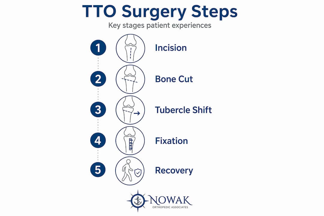
What happens during surgery
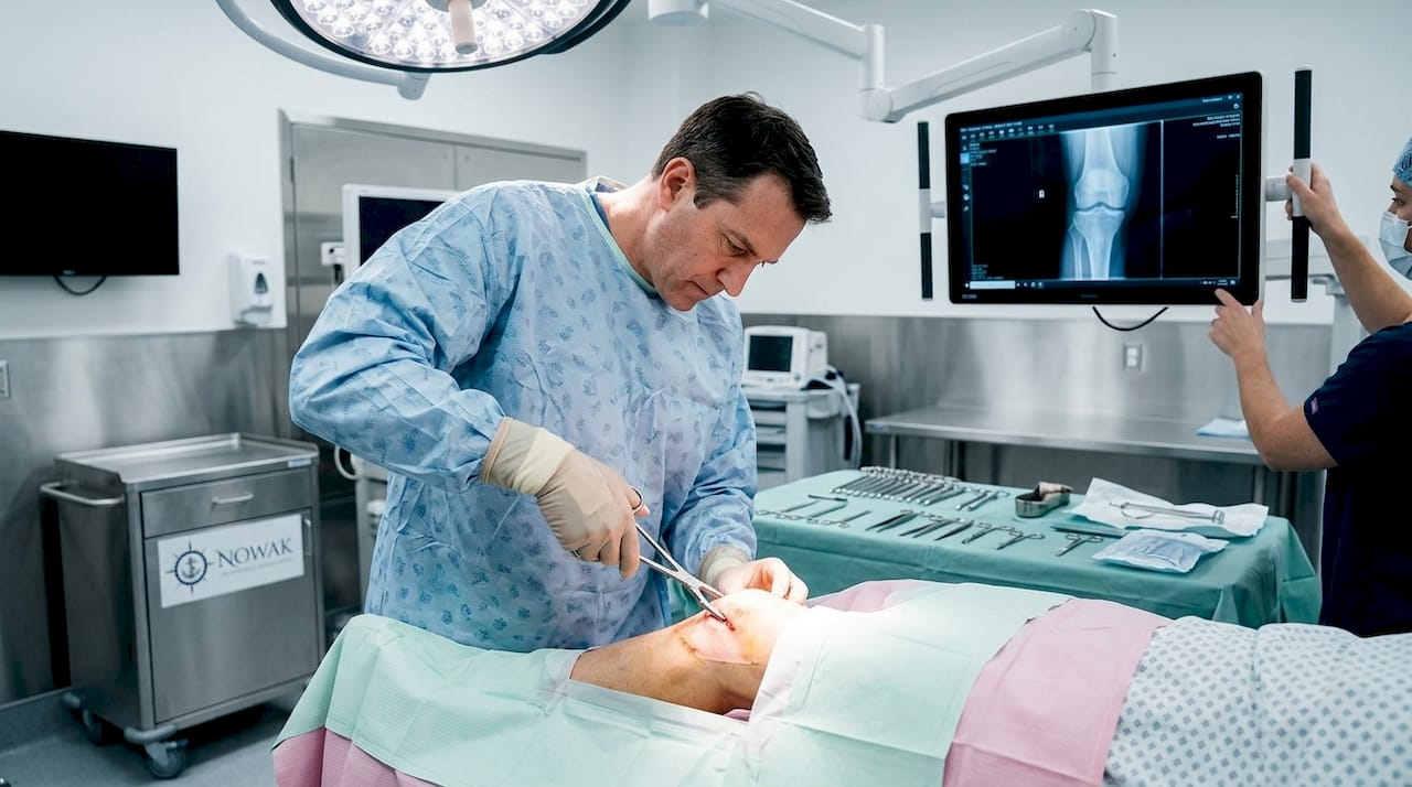
The surgeon makes an oblique cut through the tibial tubercle, then shifts the bone fragment to the planned position. Screws secure it in place. Minimally invasive techniques using arthroscopic assistance and smaller incisions are increasingly available. These approaches preserve the distal cortical periosteum, which reduces soft tissue damage and may lower complication rates compared to traditional open surgery.
Postoperative rehabilitation phases
Recovery from knee osteotomy follows a staged progression:
- Weeks 1 to 6: Partial weight-bearing with crutches; focus on controlling swelling and protecting the healing bone.
- Weeks 6 to 12: Progressive weight-bearing as bone consolidation advances; gentle range-of-motion exercises begin.
- Months 3 to 6: Strength training, proprioception work, and gradual return to functional activity.
- Months 6 and beyond: Return to sport or high-demand activity, subject to surgeon clearance.
For a detailed look at rehabilitation milestones, the post-surgery knee care guide at Nowak Orthopedic Associates walks through each phase in practical terms.
Pro Tip: Ask your surgeon about low-profile fixation hardware before the procedure. Hardware removal typically happens around 18 to 20 months post-surgery, and lower-profile screws may reduce the likelihood of irritation in the interim.
Hardware considerations
| Hardware Detail | What to Know |
|---|---|
| Type of fixation | Cortical screws most common; low-profile options available |
| When removal is needed | Average 18.7 months after TTO; most often due to anterior knee pain |
| Likelihood of removal | Approximately 21% of patients require a second procedure |
| Effect on pain | Pain scores drop an average of 3.6 points after hardware removal |
Risks, complications, and outcomes
Every surgery carries risk, and tibial tubercle osteotomy is no exception. The most frequently discussed complication is hardware irritation. Research shows that about 21% of patients require hardware removal, primarily because the screws cause anterior knee pain. The good news is that pain scores improve meaningfully after removal, dropping an average of 3.6 points on standard pain scales.
“Hardware-related reoperation after TTO is common enough that it should be part of every pre-surgical conversation, not a surprise finding during recovery.” — Pain and Hardware Removal After Tibial Tubercle Osteotomy
Notably, screw size and number do not appear to predict who will need hardware removed, which means this risk applies broadly regardless of fixation method. Other potential complications include delayed bone healing, infection, and in rare cases, the need for revision surgery. Long-term outcomes are generally positive when the correct indication is identified and surgical planning avoids overcorrection. Newer surgical systems designed for multi-directional tubercle transfer are improving precision and reducing complication rates compared to older techniques.
My perspective on TTO: what patients get wrong
I have seen tibial tubercle osteotomy evolve significantly over the years, both in technique and in how we select candidates. The most common misunderstanding I encounter is the belief that TTO is a last resort, something you only consider after everything else has failed. That framing leads patients to wait too long, often arriving with more cartilage damage than they started with.
TTO is a corrective procedure. It addresses a mechanical problem that conservative treatment simply cannot fix. Physical therapy can strengthen the muscles around a malaligned joint, but it cannot move a bone. When the anatomy is wrong, the anatomy needs to be corrected.
The second thing patients often miss is the hardware conversation. I strongly encourage anyone considering this surgery to ask their surgeon directly about fixation choices and realistic expectations for a possible second procedure. It is not a failure if hardware eventually needs to come out. It is a known and manageable part of the process, and outcomes after removal are genuinely good.
What I have learned over many years of treating patellofemoral conditions is that the patients who do best are the ones who understand their own anatomy, ask specific questions, and commit fully to rehabilitation. Surgery addresses the structure. Recovery is where function is restored.
— Dr. Roger Nowak
Ready to explore your knee treatment options?
If you are dealing with persistent knee pain, patellar instability, or have been told you may need a tibial tubercle procedure, Nowak Orthopedic Associates in Hampton, NH can help you understand your options. Dr. Roger B. Nowak specializes in advanced knee joint care, including tibial tubercle osteotomy, OATS cartilage restoration, and MPFL reconstruction. Schedule your first visit to receive a personalized evaluation and a treatment plan built around your specific anatomy and goals.
FAQ
What is tibial tubercle osteotomy used for?
Tibial tubercle osteotomy is used to correct abnormal kneecap tracking caused by conditions like patellar instability, patella alta, cartilage damage, and patellofemoral arthritis. It works by repositioning the bony attachment point of the patellar tendon to restore normal knee mechanics.
How long does recovery take after tibial tubercle surgery?
Most patients progress through rehabilitation over six months, moving from protected weight-bearing in the first six weeks to strength training and return-to-activity by months three through six. Full return to sport typically requires clearance at or after the six-month mark.
Is hardware removal after TTO normal?
Yes. Roughly 21% of patients undergo hardware removal after tibial tubercle osteotomy, most often around 18 months post-surgery due to anterior knee pain from screw irritation. Pain scores improve significantly after removal.
Can TTO be combined with other knee procedures?
Yes. TTO is frequently performed alongside MPFL reconstruction when patellar instability involves an elevated TT-TG distance, and it can be paired with cartilage restoration procedures to offload repaired tissue during healing.
How do surgeons decide which type of TTO to perform?
Surgeons use measurements like TT-TG distance and the Caton-Deschamps Index to determine whether medialization, anteriorization, distalization, or a combination is needed. The angle of the bone cut also controls how much movement occurs in each direction, which is why individualized surgical planning is so critical.