

TL;DR:
- Lateral retinacular release is a minimally invasive knee surgery that relieves pressure by cutting or lengthening tight outer knee tissue. Proper patient selection, diagnosis, and post-surgical physical therapy are essential for successful outcomes.
Lateral retinacular release is a minimally invasive knee surgery that cuts or lengthens the tight connective tissue on the outer side of the kneecap to relieve pressure and restore proper patellar tracking. If you have been dealing with persistent knee pain, clicking, or a kneecap that feels like it pulls to the outside, this procedure may be part of your treatment conversation. At Nowak Orthopedic Associates in Hampton, NH, board-certified orthopedic surgeon Dr. Roger B. Nowak performs lateral retinacular release as part of a broader approach to diagnosing and treating complex knee conditions. Understanding what this surgery involves, who qualifies, and what recovery looks like helps you make a confident, informed decision.
What is lateral retinacular release and why does it matter?
Lateral retinacular release is the surgical division or lengthening of the lateral retinaculum, a band of fibrous tissue on the outer knee that helps hold the patella in place. When this tissue becomes too tight, it pulls the kneecap outward and causes it to track improperly in the groove at the end of the femur. The result is a condition called lateral patellar compression syndrome, which produces pain at the front and outer edge of the knee, especially during activities like climbing stairs, squatting, or sitting for long periods.
A systematic review confirms that lateral retinacular release improves outcomes for patients with lateral patellar compression syndrome, anterior knee pain, and post-operative arthrofibrosis. That finding matters because it establishes this procedure as evidence-based, not experimental, when applied to the right patients.
Why does a tight lateral retinaculum cause knee problems?
The lateral retinaculum acts as a guide wire for your kneecap. When it is the right length and tension, the patella glides smoothly. When it is too tight, the kneecap tilts laterally and grinds against the outer edge of the femoral groove, wearing down cartilage and triggering inflammation.
Common symptoms of lateral retinacular tightness include:
- Anterior knee pain that worsens with stairs, squatting, or prolonged sitting
- A sensation of tightness or pulling on the outer side of the kneecap
- Crepitus, a grinding or clicking sound during knee movement
- Swelling around the kneecap after activity
- Visible lateral tilt of the patella on physical exam or imaging
Diagnosis typically combines a physical exam, where the physician assesses patellar tilt and mobility, with imaging such as X-ray or MRI. An axial view X-ray can reveal lateral patellar tilt, while MRI shows cartilage wear and soft tissue changes. These objective findings are critical because anterior knee pain has many causes, and surgery should never be the first response to pain alone.
When is lateral retinacular release recommended?
The indications for lateral retinacular release are specific. This procedure is not appropriate for every patient with knee pain, and patient selection is the single most important factor in achieving a good outcome.
Surgery is indicated only after at least 6 months of failed nonoperative treatment for significant functional impairment due to lateral patellar compression syndrome. That 6-month threshold exists because physical therapy, activity modification, bracing, and anti-inflammatory treatment resolve symptoms for many patients without any surgery at all. Exploring non-surgical orthopedic treatments first is not just a formality. It is the standard of care.
Ideal candidates share a specific profile:
- Confirmed lateral patellar compression syndrome with objective imaging evidence
- Stable kneecap with isolated lateral tilt but no generalized instability or hypermobility
- Failure of conservative care including structured physical therapy for at least 6 months
- No significant patellofemoral arthritis that would require a different surgical approach
Patients with generalized patellar instability are not good candidates. Performing release on an unstable patella can worsen instability by removing the lateral restraint without addressing the underlying alignment problem. This is why a thorough pre-surgical evaluation matters so much.
Pro Tip: Ask your surgeon specifically whether your patella is stable or unstable before agreeing to any release procedure. That single distinction changes the recommended surgery entirely.
How is the lateral retinacular release procedure performed?
The lateral retinacular release procedure is performed arthroscopically in most cases. Arthroscopic surgery uses 2 to 3 small incisions and a camera to visualize the joint, allowing the surgeon to cut or lengthen the retinaculum with precision. Arthroscopic technique results in smaller incisions, less soft tissue damage, and faster recovery compared to open surgery.

There are two distinct surgical approaches worth understanding:
| Approach | What happens | Key advantage |
|---|---|---|
| Lateral release | Retinaculum is fully cut | Faster to perform |
| Lateral lengthening | Retinaculum is lengthened via Z-plasty | Preserves structural continuity |
Lateral retinacular lengthening preserves the structural continuity of the tissue, resulting in less medial patellar instability and better outcomes than complete release. This distinction reflects a meaningful shift in surgical thinking. Many surgeons now prefer lengthening over complete release because it reduces the risk of creating a new problem on the medial side of the knee.
During surgery, the adequacy of the release is assessed by testing patellar mobility. Intraoperative success is judged by the patella tilting to neutral and gaining inward mobility. One significant intraoperative risk is bleeding from the superolateral genicular vessels, which run close to the tissue being cut. Careful technique is required to protect these vessels and reduce the risk of post-operative hematoma.
Pro Tip: When consulting with your surgeon, ask whether they plan a release or a lengthening procedure and why. The answer tells you a great deal about their familiarity with current best practices.
What is the recovery process after lateral retinacular release?
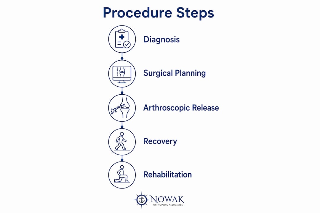
Lateral retinacular release recovery follows a structured timeline, and your commitment to rehabilitation determines your outcome as much as the surgery itself.
Most patients begin gentle range-of-motion exercises within the first few days after surgery. Structured physical therapy typically starts within one to two weeks. The goals in early recovery are reducing swelling, restoring knee flexion and extension, and preventing scar tissue from forming around the release site.
Post-operative rehabilitation is essential. Without dedicated physical therapy, scar tissue can form and effectively undo the surgical correction. This is not a minor caveat. It is the reason some patients report disappointing results after an otherwise technically successful procedure. A detailed knee surgery recovery guide can help you understand what to expect at each stage.
Key recovery milestones and considerations include:
- Weeks 1–2: Swelling management, gentle motion, and weight bearing as tolerated
- Weeks 3–6: Progressive strengthening of the quadriceps and hip stabilizers
- Weeks 6–12: Return to low-impact activity and sport-specific training
- 3–6 months: Full return to activity for most patients
Up to 20% of patients report subjective post-operative instability, though true dislocation is rare and overall symptom improvement is common with proper technique. That statistic highlights why choosing an experienced surgeon and following through with rehab are both non-negotiable parts of a good outcome.
Key Takeaways
Lateral retinacular release relieves lateral patellar compression by cutting or lengthening tight outer knee tissue, but proper patient selection and post-operative rehab determine whether surgery succeeds.
| Point | Details |
|---|---|
| Correct diagnosis first | Lateral patellar compression syndrome must be confirmed with imaging before surgery is considered. |
| Conservative care is required | At least 6 months of physical therapy must fail before surgery becomes appropriate. |
| Lengthening vs. release | Lateral lengthening preserves tissue structure and reduces the risk of medial instability. |
| Rehab drives outcomes | Skipping physical therapy after surgery risks scar tissue formation that reverses surgical gains. |
| Patient selection is critical | Patients with generalized patellar instability are not candidates and may worsen with release. |
My honest perspective on lateral retinacular release
I have performed lateral retinacular release and lengthening procedures for many years, and the most consistent lesson I have learned is this: the surgery itself is straightforward. The hard part is making sure the right patient is on the table.
Early in my career, lateral release was performed more liberally than it should have been. Patients with vague anterior knee pain and a mildly tight retinaculum were sometimes offered surgery before conservative care was truly exhausted. The results were predictably mixed. The shift toward lateral lengthening over complete release reflects a more mature understanding of patellar biomechanics, and I think it represents genuine progress.
What I tell every patient considering this procedure is that surgery corrects a structural problem. It does not fix poor quadriceps strength, poor hip mechanics, or the movement patterns that may have contributed to the problem in the first place. That is what rehabilitation addresses. Patients who invest in post-surgery knee care with the same seriousness they bring to the surgery itself consistently do better. The ones who treat physical therapy as optional rarely achieve the outcomes they hoped for.
If you are wondering whether this procedure is right for you, the answer starts with a thorough evaluation, not a quick decision.
— Dr. Roger Nowak
Explore knee care options at Nowak Orthopedic Associates
If knee pain or patellar instability has been limiting your daily activities, you do not have to keep managing it on your own. Nowak Orthopedic Associates offers a full range of orthopedic procedures for joint pain including lateral retinacular release, lateral lengthening, meniscus repair, ACL reconstruction, and more. Dr. Nowak takes time to evaluate each patient individually, confirm the correct diagnosis, and recommend the most appropriate path forward, whether that means physical therapy, a targeted injection, or a minimally invasive surgical procedure. Schedule your first visit to discuss your symptoms and explore your treatment options with a board-certified orthopedic surgeon.
FAQ
What is lateral retinacular release used to treat?
Lateral retinacular release treats lateral patellar compression syndrome, a condition where a tight outer knee ligament pulls the kneecap out of alignment and causes chronic anterior knee pain.
Who is a good candidate for this procedure?
Ideal candidates have a stable kneecap with confirmed lateral tilt, significant functional impairment, and at least 6 months of failed conservative treatment including physical therapy.
What is the difference between lateral release and lateral lengthening?
Lateral release fully cuts the retinaculum, while lateral lengthening uses a Z-plasty technique to elongate it. Lengthening preserves structural continuity and reduces the risk of medial patellar instability.
How long does lateral retinacular release recovery take?
Most patients return to low-impact activity within 6–12 weeks and reach full recovery within 3–6 months, provided they complete a structured physical therapy program.
What are the main risks of lateral retinacular release?
The primary risks include intraoperative bleeding from the superolateral genicular vessels, post-operative hematoma, and subjective patellar instability, which affects up to 20% of patients but rarely results in true dislocation.