

TL;DR:
- Most shoulder and knee pains do not require an MRI initially because physical exams and x-rays are usually sufficient. MRI is reserved for cases with failed conservative treatment, acute instability, severe injuries, or when surgery planning is necessary. Experts advise using MRI only when it will meaningfully influence treatment decisions, avoiding unnecessary scans and incidental findings.
An MRI is not the first step for most shoulder or knee pain cases. Physical examination and x-rays are the standard starting point, covering over 90% of non-traumatic cases before advanced imaging is ever considered. The American College of Radiology (ACR) Appropriateness Criteria reserve MRI for situations where the result will actually change how a patient is treated. At Nowak Orthopedic Associates, the decision to order an MRI follows that same evidence-based logic: imaging should answer a clinical question, not just confirm that pain exists.
Do I need an MRI for shoulder or knee pain right away?
The short answer is: probably not at first. Physical exams and x-rays are more sensitive and specific than most patients expect, and they guide the majority of orthopedic diagnoses without any advanced imaging.
MRI becomes appropriate when specific clinical triggers are present. The ACR guidelines and orthopedic practice both point to the same key thresholds:
- Failed conservative treatment: If physical therapy, anti-inflammatory medication, or activity modification has not improved symptoms after 4–8 weeks, MRI becomes a reasonable next step.
- Acute instability or locking: A knee that locks mid-stride or a shoulder that repeatedly dislocates signals possible structural damage that warrants early imaging.
- Inability to bear weight: This symptom after an acute injury can indicate a fracture, ligament rupture, or severe meniscus tear requiring prompt evaluation.
- Surgical planning: When an orthopedic surgeon needs to confirm the extent of a tear or plan a procedure, MRI provides the detail that x-rays cannot.
- Inconclusive clinical picture: When the physical exam and x-rays do not explain the severity of symptoms, MRI helps clarify the diagnosis.
Chronic pain and acute injury follow different pathways. Acute trauma with clear clinical signs may justify faster MRI access. Chronic joint pain almost always starts with conservative care first.
Pro Tip: Keep a written record of your physical therapy sessions and symptom changes. Documented conservative treatment failure is the primary factor insurance companies use to approve an MRI.

How do physical exams and x-rays compare to MRI?
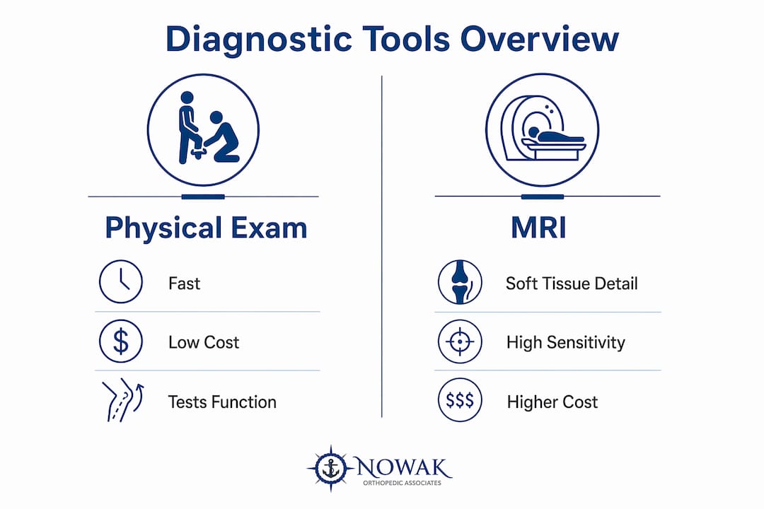
A thorough physical examination by an orthopedic specialist remains the most direct diagnostic tool available. Clinical evaluation often provides sufficient answers without any imaging at all. Specific provocative tests, range-of-motion measurements, and strength assessments can identify rotator cuff weakness, meniscus irritation, and ligament laxity with high accuracy.
X-rays excel at detecting fractures, bone spurs, joint space narrowing from arthritis, and alignment problems. They are fast, low-cost, and widely available. Weight-bearing x-rays of the knee, in particular, give a realistic picture of how the joint functions under load.
MRI adds soft tissue detail that x-rays cannot provide. It is highly sensitive for rotator cuff tears, reaching 96.5% sensitivity in observational studies. That said, ultrasound shows comparable diagnostic accuracy for many common shoulder pathologies at a fraction of the cost and without the wait time.
| Diagnostic tool | Strengths | Limitations |
|---|---|---|
| Physical exam | Fast, low-cost, tests function directly | Operator-dependent; limited for deep structures |
| X-ray | Detects fractures, arthritis, alignment | Cannot visualize soft tissue |
| Ultrasound | Real-time soft tissue imaging, low cost | Limited for deep joint structures |
| MRI | Best soft tissue detail, high sensitivity | Expensive, slow, incidental findings common |
Pro Tip: Ask your orthopedist whether ultrasound could answer the same clinical question as an MRI. For rotator cuff assessment, it often can, and it is faster to schedule.
What are common misconceptions about MRI for joint pain?
The biggest misconception is that MRI is the definitive test for joint pain. Patients commonly believe that a clinical evaluation cannot provide sufficient answers, but that belief underestimates what a skilled orthopedic exam can reveal.
A second misconception is that a positive MRI finding explains all symptoms. MRI can detect incidental findings unrelated to the patient’s actual pain, including minor tears and anatomical variations that would never cause problems. Without experienced clinical interpretation, these findings can trigger unnecessary anxiety or even inappropriate surgery.
“Imaging findings must always be correlated with the patient’s symptoms and physical examination. An MRI that shows a small labral fraying in a 50-year-old may be completely incidental. Treating the image rather than the patient is one of the most common pitfalls in orthopedic care.”
A third misconception involves insurance. Insurance approval typically requires documented failure of conservative management before an MRI is authorized, except in cases of acute trauma with clear clinical signs. Patients who skip physical therapy and go straight to requesting an MRI often face delays and denials.
The practical takeaway: MRI is most useful when it will change the treatment plan. If the result will not alter what happens next, the scan adds cost and complexity without benefit.
When should you consider an MRI and how does it change treatment?
MRI earns its place in orthopedic care when the clinical stakes are high enough to justify it. Here are the scenarios where it genuinely shifts treatment decisions:
- Surgical evaluation for meniscus tears. When a patient presents with joint line tenderness, clicking, and failed conservative care, MRI confirms the tear pattern and guides whether meniscus repair or resection is appropriate.
- Rotator cuff injury assessment. A full-thickness rotator cuff tear requires different management than a partial tear. MRI defines the size and retraction of the tear, which directly influences whether rotator cuff repair surgery is indicated.
- ACL and ligament damage. After a significant knee injury with instability, MRI identifies complete versus partial ACL tears and checks for concurrent meniscus or cartilage damage before ACL reconstruction is planned.
- Cartilage restoration planning. Procedures like the osteochondral autograft transfer system (OATS) require precise mapping of cartilage defects. MRI provides that map before surgery.
- Complex or ambiguous cases. When symptoms are severe but x-rays are normal and the physical exam is inconclusive, MRI resolves the uncertainty and prevents both undertreatment and overtreatment.
Ordering the correct MRI protocol matters as much as ordering the scan itself. A generalized MRI without targeted protocols reduces diagnostic usefulness. At Nowak Orthopedic Associates, the specific protocol, including whether contrast is used, is selected based on the suspected pathology before the order is placed.
Patients considering non-surgical options such as platelet-rich plasma (PRP) injections or SoftWave therapy may also benefit from MRI to confirm the diagnosis and target treatment accurately. For patients exploring light therapy for joint injuries as a complementary approach, imaging context still helps guide where and how treatment is applied.
Key Takeaways
MRI for shoulder or knee pain is appropriate only when it will change the treatment plan, not as a routine first step.
| Point | Details |
|---|---|
| Start with exam and x-ray | Physical exams and x-rays cover the majority of joint pain diagnoses before MRI is needed. |
| MRI follows failed conservative care | After 4–8 weeks without improvement, MRI becomes clinically and insurance-justified. |
| Incidental findings are real risks | MRI can show abnormalities unrelated to pain; expert interpretation prevents overtreatment. |
| Protocol selection matters | Ordering the right MRI type for the suspected condition produces far more useful results. |
| MRI drives surgical decisions | Its greatest value is confirming surgical candidacy and planning procedures like ACL reconstruction or rotator cuff repair. |
My take on when MRI actually earns its place
Patients often arrive expecting an MRI to be the answer. I understand that impulse. When you are in pain, you want certainty, and a scan feels like certainty. But in my experience, the physical examination tells me more than most patients expect, and ordering an MRI before I know what question it is answering rarely helps anyone.
The cases where I find MRI indispensable are the ones where surgery is genuinely on the table. If I am planning a rotator cuff repair, a meniscus repair, or an ACL reconstruction, I need that soft tissue detail. I need to know the tear size, the tissue quality, and what else might be involved. That is when MRI changes what I do in the operating room.
What concerns me more is the MRI ordered out of habit or patient pressure, without a clear clinical question behind it. Those scans come back with findings that are real on the image but irrelevant to the pain, and then I spend the appointment reassuring a patient that the “tear” we found is not the problem. That is a cost, in time, money, and anxiety, that serves no one.
My advice: trust the process. A thorough shoulder injury evaluation or knee assessment by a board-certified orthopedic surgeon is the right starting point. If MRI will help you, you will know why, and it will be ordered with purpose.
— Dr. Roger Nowak
Orthopedic care at Nowak Orthopedic Associates: from evaluation to imaging
At Nowak Orthopedic Associates in Hampton, NH, every patient begins with a thorough clinical evaluation before any imaging decision is made. Dr. Roger B. Nowak uses a structured, evidence-based approach to determine whether x-rays, ultrasound, or MRI is the right next step for your specific condition. When MRI is indicated, the correct protocol is selected from the start, so the scan answers the right question. Whether you are dealing with a suspected meniscus tear, rotator cuff injury, or persistent joint pain that has not responded to conservative care, the team provides advanced joint care built around your clinical picture. Schedule your first visit to get a clear diagnosis and a treatment plan grounded in what your body actually needs.
FAQ
Is an MRI always needed to diagnose a torn meniscus?
No. A physical exam with specific provocative tests can strongly suggest a meniscus tear. MRI confirms the diagnosis and defines the tear pattern when surgery is being considered.
How long does conservative treatment need to fail before insurance approves an MRI?
Most insurers require a documented course of 4–8 weeks of conservative treatment, such as physical therapy, before approving an MRI for non-traumatic joint pain.
Can an MRI show something that is not actually causing my pain?
Yes. MRI can detect incidental anatomical findings like minor tears or variations that are unrelated to symptoms. Expert clinical correlation is required to avoid unnecessary treatment.
Is ultrasound a reasonable alternative to MRI for shoulder pain?
For many shoulder conditions, yes. Ultrasound shows comparable diagnostic accuracy to MRI for common pathologies like rotator cuff tears and is faster and less expensive to obtain.
When does an orthopedic surgeon order an MRI before trying conservative treatment?
Early MRI is appropriate when acute symptoms include joint locking, severe instability, or inability to bear weight, as these signs suggest structural damage that may require prompt surgical evaluation.